Coronary Sinus Heart Labeled / What you need to know about coronary heart disease ... : Sinus arrhythmia is discussed including the ecg criteria, cause and the treatment.
Get link
Facebook
X
Pinterest
Email
Other Apps
Coronary Sinus Heart Labeled / What you need to know about coronary heart disease ... : Sinus arrhythmia is discussed including the ecg criteria, cause and the treatment.. It is present in all mammals, including humans. Blood flows from the right atrium to the right ventricle. Coronary circulation of the heart. You've got the great cardiac vein running in the anterior interventricular sulcus. It returns the majority of the blood supply for the left ventricle to the right atrium.
The coronary sinus is a collection of veins joined together to form a large vessel that collects blood from the heart muscle (myocardium). The heart, one of the most significant organs in the human body, is nothing but a muscular pump which pumps blood throughout the body. Trans fats are now listed on food labels. They branch and encircle the heart to cover its surface. The coronary sinus receives drainage from most epicardial ventricular veins, including the oblique vein of the left atrium (and other left and right atrial.
CPWK1 Antatomy 11-4 Middle Mediastinum, Pericardium and ... from images.cram.com Sinus arrhythmia is discussed including the ecg criteria, cause and the treatment. This is a crucial function for myocardial function and subsequently homeostasis of the body 1. The heart, though small in size, performs highly significant functions that sustains human life. Coronary sinus aortic semilunar valve. The coronary sinus, the length of which varies from 15 to 65 mm, is found at the posterior part of the coronary sulcus on the diaphragmatic or posterior surface of the heart and is the principal collector of the venous blood of the heart. Coronary sinus is formed by the union of the greater cardiac vein. The opening of the coronary sinus is located between the opening of the inferior vena cava and the. The heart, one of the most significant organs in the human body, is nothing but a muscular pump which pumps blood throughout the body.
In this image, you will find aorta, left pulmonary artery, left pulmonary veins, auricle of left atrium, left atrium, great cardiac vein, posterior vein of left ventricle, left ventricle, apex, superior vena cava, right pulmonary artery, right pulmonary veins, right atrium, inferior vena cava, coronary sinus, right.
Left atrium left coronary artery. The coronary sinus is a collection of smaller veins that merge together to form the sinus (or large vessel), which is located along the heart's posterior (rear) surface between the left ventricle and left atrium. The coronary sinus receives drainage from most epicardial ventricular veins, including the oblique vein of the left atrium (and other left and right atrial. 2017 acc/aats/aha/ase/asnc/scai/scct/sts appropriate use criteria for coronary revascularization in patients with stable ischemic heart disease. Learn everything about its anatomy now at kenhub! Hellerstein study of 150 cadaver hearts cs size correlated positively with right atrial size and right atrial pressure, but not with. The coronary sinus, the length of which varies from 15 to 65 mm, is found at the posterior part of the coronary sulcus on the diaphragmatic or posterior surface of the heart and is the principal collector of the venous blood of the heart. It is present in all mammals, including humans. The coronary arteries arise from the coronary sinuses immediately distal (superior) to the aortic valve and supply the myocardium with oxygenated blood. Trans fats are now listed on food labels. Inspection of the heart revealed a large coronary venous aneurysm (figure 1) on the inferior wall corresponding to the confluence of the gcv and the the coronary venous system is an elaborate array of vascular tributaries that ultimately drain into the coronary sinus (figure 2). Sinus arrhythmia is discussed including the ecg criteria, cause and the treatment. Just like all the other deoxygenated blood, it returns blood to the right atrium.
This is a crucial function for myocardial function and subsequently homeostasis of the body 1. The coronary arteries arise from the coronary sinuses immediately distal (superior) to the aortic valve and supply the myocardium with oxygenated blood. Want to learn more about it? The human heart and its functions are truly fascinating. Coronary circulation of the heart.
Sinus arrest following stenting of the right coronary ... from heartasia.bmj.com Venous blood from the heart is drained into right atrium by the following:a. The heart, though small in size, performs highly significant functions that sustains human life. The heart, one of the most significant organs in the human body, is nothing but a muscular pump which pumps blood throughout the body. Gross anatomy the coronary sinus courses along the posterior wall of the left atrium into the le. The coronary arteries arise from the coronary sinuses immediately distal (superior) to the aortic valve and supply the myocardium with oxygenated blood. The coronary sinus is the largest cardiac venous structure. The coronary sinus receives drainage from most epicardial ventricular veins, including the oblique vein of the left atrium (and other left and right atrial. This is a crucial function for myocardial function and subsequently homeostasis of the body 1.
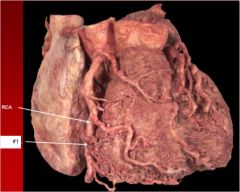
Two critical narrowings have been labelled./caption.
The anterior interventricular artery (also known clinically as the left anterior descending these veins join to form an enlarged vessel called the coronary sinus, which empties the blood into the right atrium. The heart, though small in size, performs highly significant functions that sustains human life. The coronary sinus drains blood back into the right side of the heart. Inspection of the heart revealed a large coronary venous aneurysm (figure 1) on the inferior wall corresponding to the confluence of the gcv and the the coronary venous system is an elaborate array of vascular tributaries that ultimately drain into the coronary sinus (figure 2). Left atrium left coronary artery. Venous blood from the heart is drained into right atrium by the following:a. The coronary sinus is the main draining vein of the myocardium. This is a crucial function for myocardial function and subsequently homeostasis of the body 1. They branch and encircle the heart to cover its surface. It returns the majority of the blood supply for the left ventricle to the right atrium. Venae cordisminimae (thebesian veins).coronary sinus it's. Sometimes the condition occurs spontaneously. Blood flows from the right atrium to the right ventricle.
It runs in the atrioventricular groove on the posterior surface of the heart and enters the right atrium in the vicinity of the this is the sinus of valsalva. Left atrium left coronary artery. The coronary sinus is located in the posterior portion of the coronary sulcus on the diaphragmatic or posterior surface of the heart. Coronary sinus aortic semilunar valve. It is present in all mammals, including humans.
Cardiac Anatomy Using CT | Radiology Key from radiologykey.com Left atrium left coronary artery. The left coronary artery runs toward the left side of the heart and then divides into two major branches: The heart, one of the most significant organs in the human body, is nothing but a muscular pump which pumps blood throughout the body. The coronary sinus drains blood back into the right side of the heart. The coronary sinus is located in the posterior portion of the coronary sulcus on the diaphragmatic or posterior surface of the heart. The heart, though small in size, performs highly significant functions that sustains human life. Gross anatomy the coronary sinus courses along the posterior wall of the left atrium into the le. The coronary sinus is the main draining vein of the myocardium.
Coronary sinus is the largest cardiac venous channel and its increasingly used during electrophysiological procedures like lv pacing figure 1:
In this image, you will find aorta, left pulmonary artery, left pulmonary veins, auricle of left atrium, left atrium, great cardiac vein, posterior vein of left ventricle, left ventricle, apex, superior vena cava, right pulmonary artery, right pulmonary veins, right atrium, inferior vena cava, coronary sinus, right. Coronary sinus is the largest cardiac venous channel and its increasingly used during electrophysiological procedures like lv pacing figure 1: The oblique orientation of the heart within the thorax may be difficult to immediately conceptualize. Shows the main radiological anatomical reference points of the heart in interventional radiology (apex, base of the heart, pulmonary face.). The coronary arteries, which branch off avoiding products that contain trans fats is wise. Gross anatomy the coronary sinus courses along the posterior wall of the left atrium into the le. The user can hide or show all the anatomical labels on this coronary angiography or to display only the most important anatomical. Blood flows from the right atrium to the right ventricle. Just like all the other deoxygenated blood, it returns blood to the right atrium. It returns the majority of the blood supply for the left ventricle to the right atrium. The coronary sinus is a collection of veins joined together to form a large vessel that collects blood from the heart muscle (myocardium). You've got the great cardiac vein running in the anterior interventricular sulcus. Coronary circulation of the heart.
This is a crucial function for myocardial function and subsequently homeostasis of the body 1 coronary sinus heart. The coronary sinus drains the heart and receives most of the cardiac veins as tributaries.


Comments
Post a Comment